


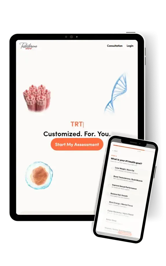
Consult Licensed Providers. Start Treatment From home.
Testosterone
Nandrolone Decanoate, hCG
Oxandrolone, Cypionate,
Enanthate
Anti-Aging
Vitamins & Amino Acids
HGH
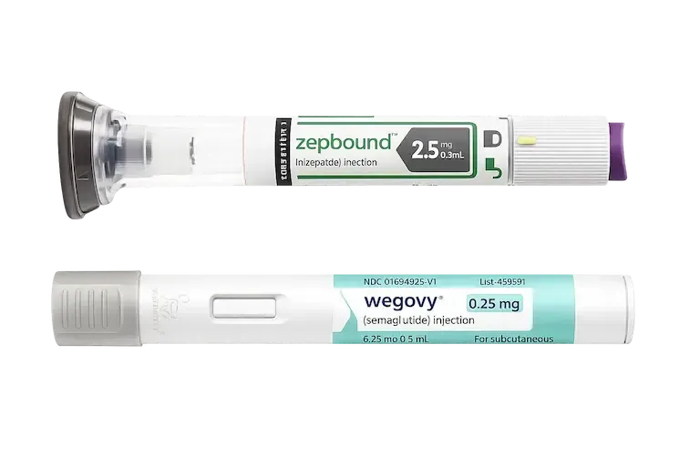
Weight Loss
Wegovy® & Semaglutide
Zepbound® & Tirzepatide
Brain Focus
Unlock Mental Clarity
Modafinil & Limitless Drug

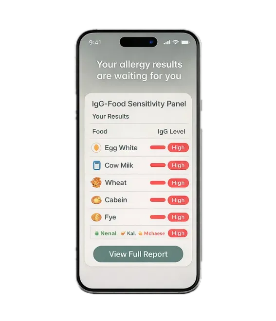
Food & Gut Allergies
Discover What's Irritating
Your Gut with an At-Home Test
Detox
Antiparasitics & Antibiotics
Intestinal & Liver Cleanse

Hair Restoration
Repair Scalp Damage
Vascularize Hair Follicles
Sleep
Medical Sleep Support for
Deeper, More Consistent Rest
Skincare
Target Wrinkles, Rosacea,
Eczema, & Acne Scars
Blood Work
Check Hormones, Allergies, Biological Age, & Organ Health
Sexual Health
Sildenafil, Tadalafil, Oxytocin,
PT-141. Fast-Acting

Supplements
20% off Top Clinical Brands:
Thorne® Pure® Metagenics®


Interested in Prescription Peptides?
Peptides are short chains of amino acids that signal your body to repair, recover, and perform. Learn the science behind them.

Interested in Prescription Peptides?
Peptides are short chains of amino acids that signal your body to repair, recover, and perform. Learn the science behind them.
Explore by Rx Treatment
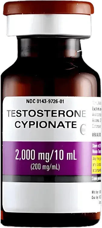
Testosterone & hCG
Testosterone Cypionate & Pregnyl (hCG)
Gold Standard for TRT Optimization
✅ Increase total testosterone levels
✅ Maintain natural sperm production
✅ Improve energy, libido, & mood
includes Pregnyl (hCG)
Consultation & blood work required
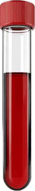
TRT Blood Work
Baseline labs for TRT diagnosis:
Total Testosterone, PSA, CBC
✅ Quest Diagnostics
✅ Prostate screening
✅ Assess Liver & Kidney function
Lab order and results review included
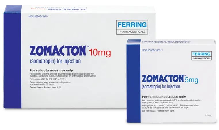
Zomacton®
FDA-approved somatropin
Growth Hormone Deficiency
✅ Restores GH-IGF-1 axis
✅ Supports lean mass & bone mineral density
✅ Regulates protein synthesis + cellular turnover
Consultation & blood work required

Oxandrolone
DHT-derived anabolic that binds
androgen receptors
✅ Increases bone mineral density
✅ Preserves lean tissue
✅ Supports nitrogen retention
Consultation & blood work required
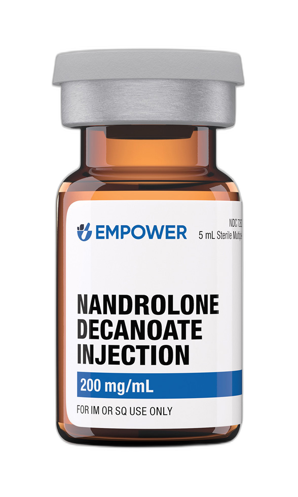
Nandrolone Decanoate
19-nortestosterone derivative that binds
androgen receptors
✅ Indicated for anemia in renal insufficiency
✅ Supports protein synthesis
✅ Preserves lean tissue in caloric deficiency
Consultation & blood work required
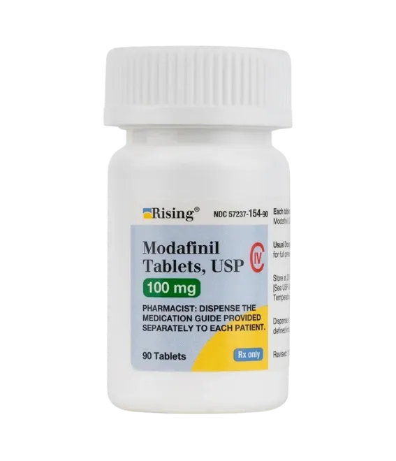
Modafinil
Wakefulness-promoting nootropic that
enhances cognitive performance
✅ Razor-sharp focus
✅ No jitters, no crash
✅ Unlock peak mental performance
Consultation & care coordination included
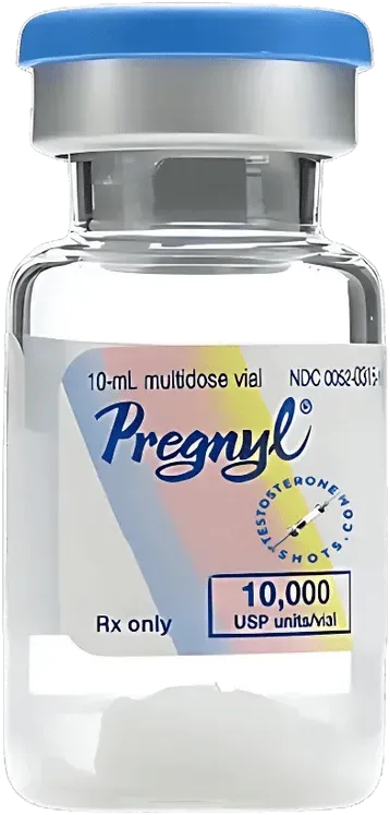
Pregnyl (hCG)
FDA-approved brand-name human chorionic gonadotropin
✅ Mimics luteinizing hormone (LH)
✅ Increase sperm quantity & quality
✅ Supports testosterone production
Consultation & care coordination included

T-Booster
4 in 1: Enclomiphene + DHEA
Pregnenolone + Zinc
✅ Boost natural testosterone
✅ Balance hormones
✅ Increase energy, libido, & mood
Consultation & care coordination included
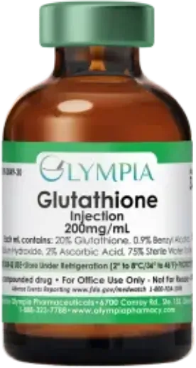
Glutathione
Reduces intestinal oxidative stress
and supports liver detox
✅ Protects gut lining
✅ Brighter, clearer skin tone
✅ Boosts immune defense and recovery
Consultation & care coordination included

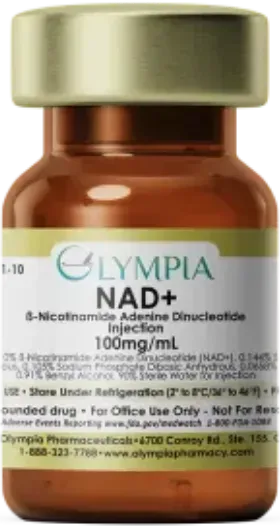
NAD+
Fuels cellular energy production & DNA
repair in every cell
✅ Recharge your cells from within
✅ Sharper focus, sustained energy
✅ Slow aging at the cellular level
Consultation & care coordination included
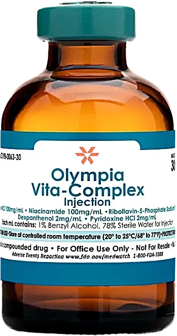
Vitamin B-Complex
Vitamins: Thiamine + Riboflavin + Niacinamide
Dexpanthenol + Pyridoxine
✅ Natural energy & reduced fatigue
✅ Eliminate brain fog & improve mood
✅ Healthier hair, skin, & nails
Consultation & care coordination included
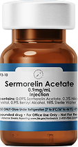
Sermorelin
Synthetic growth hormone-releasing peptide
that stimulates the pituitary gland
✅ Deeper sleep & faster recovery
✅ Improved energy & lean muscle
✅ Supports joint health
Consultation & care coordination included

Gooning
Tadalafil + Oxytocin + PT-141
Ultimate Performance
✅ Tadalafil for erections
✅ Oxytocin for sexual desire
✅ PT-141 for horniness
Consultation & care coordination included
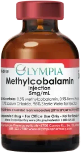
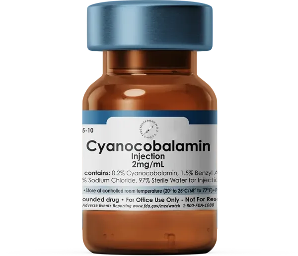
Vitamin B12
Methylcobalamin, the bioactive form of B12
✅ Boosts energy & reduces fatigue
✅ Supports brain clarity
✅ Promotes red blood cell production
Consultation & care coordination included
NAD+ Sublingual
Vitamin B3-derived coenzyme
Boosts mitochondrial ATP production
✅ Sustained cellular energy & longevity
✅ Detox & DNA repair
✅ Improved cognitive clarity & mood
Consultation & care coordination included
Stella +
4-in-1: Estriol + GHK-Cu
Niacinamide + Tretinoin
✅ Reverse fine lines and wrinkles
✅ Restore collagen and skin elasticity
✅ Achieve brighter & firmer skin
Consultation & care coordination included
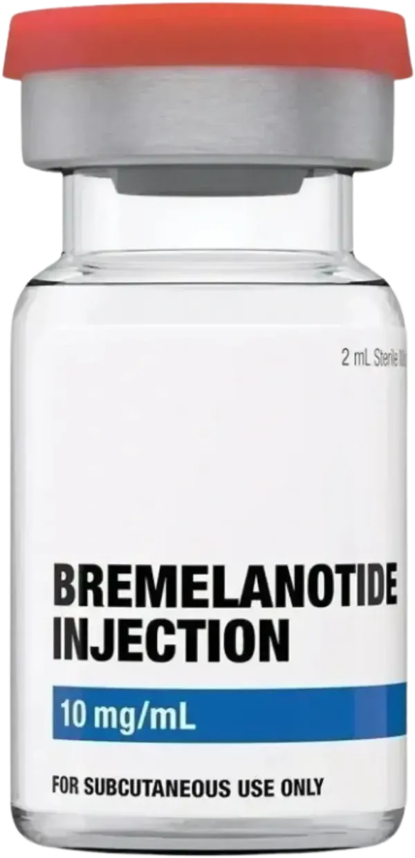
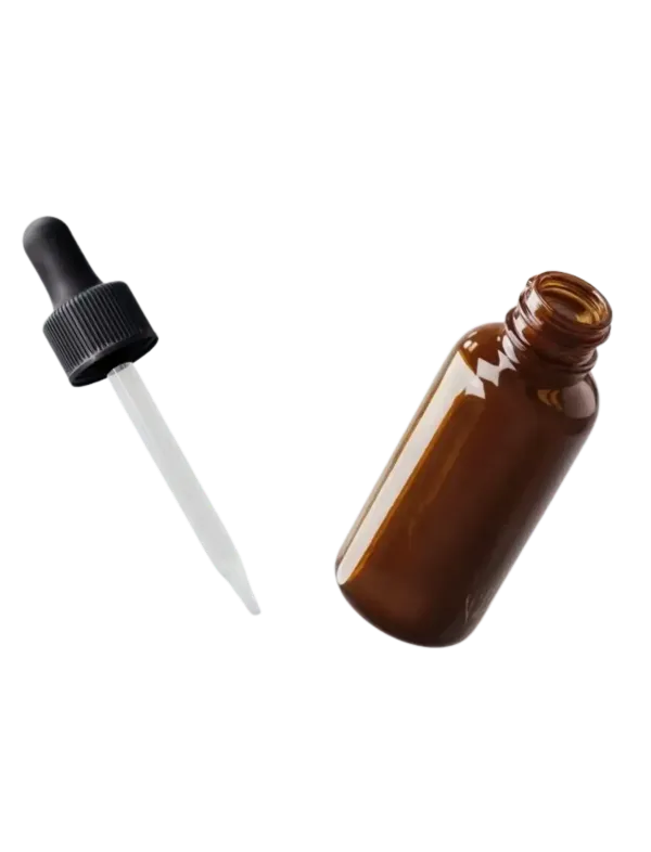
PT-141
Sexual desire & arousal activated via central nervous system
✅ Boost horniness and sensitivity
✅ Works for men and women
✅ Effects last up to 72 hours
Consultation & care coordination included
Minoxidil + Finasteride
Finasteride blocks DHT
Minoxidil stimulates regrowth
✅ Stop thinning at the root cause
✅ Visible regrowth
✅ Two proven ingredients, one protocol
Consultation & care coordination included

Food & Gut Allergy
Hidden triggers behind bloating, fatigue,
brain fog, joint pain, and skin flares that show
up hours or days after eating
✅ At-home finger-prick kit
✅ Meat, Fish, Dairy, Eggs, Nuts, Seeds, Oils
✅ Vegetables, Legumes, Fruit, Grain, Spices
Includes provider order & results review
Tadalafil Sublingual
Dissolved under the tongue for faster absorption
✅ Absorbs in minutes, not hours
✅ Lasts up to 36 hours
✅ Be ready when the moment hits
Consultation & care coordination included
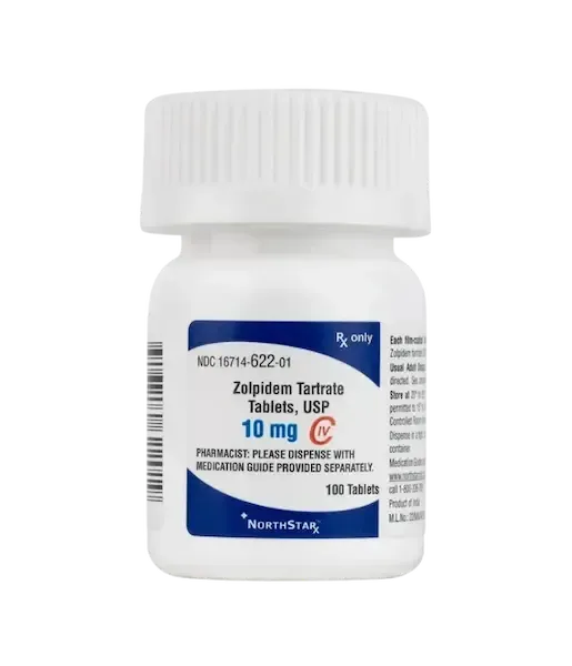
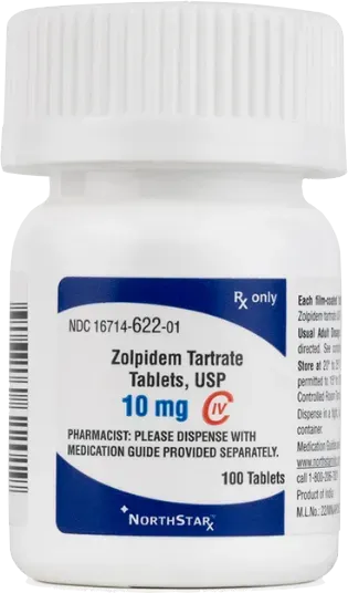
Zolpidem
Non-benzodiazepine sedative binds to GABA
receptors for rapid sleep onset
✅ Fall asleep in 15 minutes
✅ Full night of restorative sleep
✅ Wake refreshed, no grogginess
Consultation & care coordination included
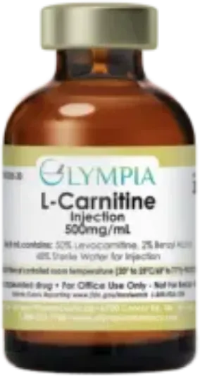
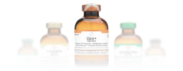
Carnitine
Shuttles fatty acids into mitochondria
for energy and fat oxidation
✅ Turn stored fat into fuel
✅ Enhance endurance and recovery
✅ Upregulate androgen receptor density
Consultation & care coordination included
Enclomiphene
Selective estrogen receptor modulator that
signals your brain to produce more LH & FSH
✅ Raise testosterone without injections
✅ Protect sperm count and fertility
✅ Oral pill, taken once daily
Consultation & care coordination included

Environmental Allergy
Discover immediate allergy-triggering
symptoms like hives, skin flushing, eye redness,
swelling, & respiratory irritation
✅ Trees, Grass, Weeds, Mold, Fungus
✅ Pet Dander, Dust Mite, Insect, Pollen
✅ At-home finger-prick kit
Includes provider order & results review
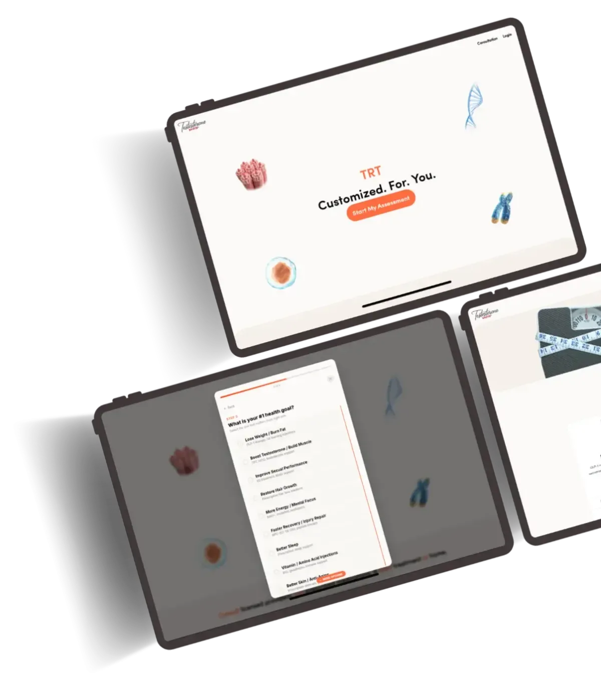
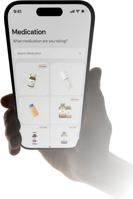
How It Works
Choose a treatment
Browse a curated selection of medically guided treatments designed for real needs, not trends. Each option is clearly explained so you can make an informed choice without medical jargon or confusion.
Complete a secure medical intake
Fill out a confidential health questionnaire that helps our providers understand your medical history,
goals, and current condition. Your information is encrypted and reviewed with strict privacy standards.
Provider review & approval
A licensed medical provider reviews your intake to ensure the treatment is personalized to your goals. They may request additional details or lab work before approval.

Prescription Sent to Pharmacy
Once approved, your prescription is sent to a licensed pharmacy for fulfillment. The pharmacy will prepare and ship your medication discreetly to you, fitting seamlessly into your routine.
Private. Simple. Delivered.
100% Online Medical
Receive professional medical care entirely online, from consultation to prescription management. No waiting rooms, no appointments to commute to - everything happens on your schedule.
Pharmacy Shipping

Your privacy comes first. All medications are shipped in plain, unbranded packaging with no indication of contents, ensuring a confidential and stress-free delivery experience.
Ongoing Support
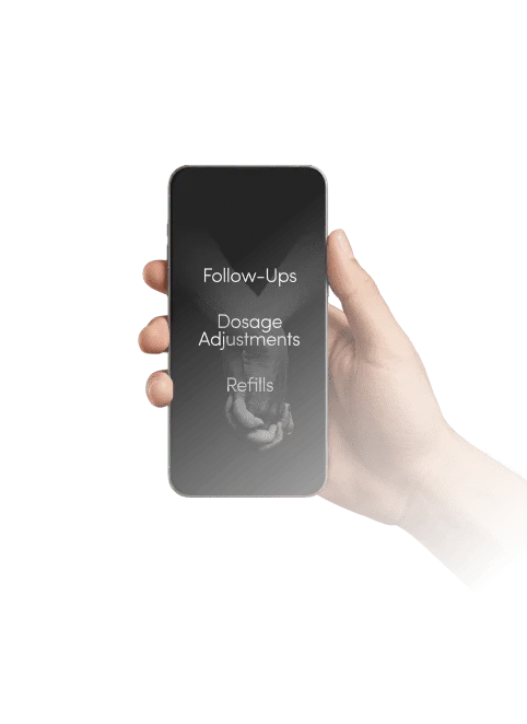
Care doesn’t stop once you receive your medication. Stay connected with licensed providers for follow-ups, dosage adjustments, refills, and questions through your secure patient portal.
Trust & Compliance
HIPAA-Compliant Platform
We use secure, HIPAA-compliant systems to protect your personal and medical data at every step, from intake forms to ongoing care. Your information is never shared without authorization.
Licensed Medical Providers
All treatments are reviewed and approved by licensed medical professionals who follow
established clinical guidelines and regulatory requirements.
Partner Licensed Pharmacies
Prescriptions are fulfilled by licensed 503A or 503B compounding pharmacies that meet strict quality, safety, and compliance standards.
Proof, Not Promises
High Standards
Higher Ratings

Our Experts

Dr. Gideon Kwok, DO
Medical Director
Ahmed Mahdi, DNP
Practitioner
Our Partners


FAQs
Are you a pharmacy?
Testosterone Shots is a telehealth platform, not a pharmacy. We provide online consultations, clinical review, and care coordination. If treatment is approved, your prescription is sent to a licensed pharmacy partner, who handles medication payment, fulfillment, and shipping directly to you.
Do I need blood work?
Our medical providers recommend a baseline lab panel for a personalized treatment plan based on your biomarkers. This helps us understand your unique health profile, prescribe the right treatment at the right dose, and monitor your progress over time. We offer 16 lab panels starting at $99 through Quest Diagnostics, with results in 3-5 business days.
How does the process work?
Complete a secure medical intake form and consult with a licensed provider through telehealth. If treatment is approved, your prescription may be sent to a licensed pharmacy partner. The pharmacy contacts you directly to verify shipping information, verify billing information, and collect payment for the medication. The pharmacy then dispenses and ships the medication directly to you, where permitted by law.
How long does it take to get my medication?
After provider approval, timing depends on the pharmacy. Most patients receive pharmacy-shipped medications within 5–7 business days after the pharmacy completes payment and shipping confirmation.
When will I start feeling results?
Results depend on the treatment and individual health profile. Every patient's experience is different and results may vary. Your provider will discuss realistic expectations during your consultation and adjust your treatment plan based on your progress and lab work.
Who will I see for my consultation?
All consultations are conducted by licensed medical providers, including DNPs, MDs, or DOs, who specialize in each treatment area.
Do you accept insurance?
We do not accept insurance at this time. Patients may be able to use insurance at Quest Diagnostics or Labcorp to help cover the cost of blood work.
What if I'm not approved for treatment?
Our goal is to help you become healthy. Under the guidance of our medical providers, we personalize a treatment plan tailored to your goals, biomarkers, and DNA. If a specific treatment isn't right for you, a provider will recommend alternative options to help you reach your health goals.
What is your refund policy?
Consultation, clinical review, care coordination, membership, and lab-related fees may be subject to the refund terms listed in our Terms of Service. Testosterone Shots does not collect payment for prescription medication and does not dispense, ship, or accept returns of prescription medication. Medication billing, payment, dispensing, shipping, returns, and refund issues are handled directly by the dispensing pharmacy, subject to pharmacy policy and applicable law.
Contact Us
Start Your Journey
Finally Feel Like
Yourself Again
Our doctors listen first.
Licensed Provider
Licensed Pharmacy
HIPAA Compliant
LegitScript

© 2026 TestosteroneShots.com, PC
text (323)-283-9219
638 1/2 N. Robertson Blvd, West Hollywood, CA 90069
Legal
The information and clinical services described on this website are for educational and informational purposes only and are not intended to diagnose, treat, cure, or prevent any disease. All treatments require evaluation and approval by a licensed healthcare provider through a telemedicine consultation. Treatment approval is not guaranteed. Individual results may vary, and treatments may carry risks and side effects. Certain compounded medications, wellness treatments, or off-label uses may not be evaluated by the U.S. Food and Drug Administration (FDA) for safety, effectiveness, or quality unless explicitly stated. Testosterone Shots provides telehealth consultations, clinical review, care coordination, lab-related services, membership services, and ongoing treatment management. Testosterone Shots is not a pharmacy and does not manufacture, compound, dispense, sell, handle, warehouse, ship, or collect payment for prescription medication. Testosterone Shots collects payment only for consultations, clinical review, care coordination, lab-related services, membership services, and other non-pharmacy services. If treatment is approved, your prescription may be sent to a licensed pharmacy partner. The pharmacy contacts you directly to complete medication payment and shipping. Services are provided by licensed physicians, nurse practitioners, and/or other licensed healthcare providers in states where they are authorized to practice. This service is not intended for medical emergencies. If you are experiencing an emergency, call 911 or seek immediate medical care. We take reasonable measures to protect personal health information in accordance with applicable privacy laws, including HIPAA. By using this website or its services, you agree to our Terms of Service, Privacy Policy, Notice of Privacy Practices, Telehealth Consent, and Important Safety Information. You must be 18 years of age or older to use this service.